In complex biological systems, critical information is often hidden beneath the surface. For Dr Maria Merin Antony, an NTU MAE research fellow from The Centre for Optical and Laser Engineering (COLE), the challenge is to reveal this hidden information without physically altering the system.
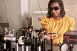
 © Nanyang Technological University SingaporeDr Maria in the lab working on the system
The limits of what can be seen
Conventional imaging systems are constrained by what the human eye can perceive. Standard cameras capture only three colour channels, red, green and blue, which provide a simplified view of reality.
Hyperspectral imaging expands this view by recording hundreds of spectral bands. This allows materials to be identified through their unique spectral signatures, making it possible to detect subtle variations in composition, structure and condition.
This capability is particularly valuable in biological systems, where critical information often lies beneath surface layers. Structures such as plant leaves and the human cornea are optically complex, consisting of multiple layers that interact with light in different ways. Accessing this information without invasive techniques has long been a challenge.
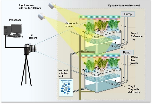 © Nanyang Technological University SingaporeFigure 1: Schematic diagram of the Hyperspectral Imaging (HSI) based monitoring platform for hydroponic lettuce. The blue dashed line indicates the field of vision of the lens used.
A breakthrough in deep-layer imaging
Dr Maria's work addresses this limitation by combining optical system design with advanced computational analysis. To demonstrate the technology, she studied two representative biological models: plant leaves and the human cornea, both of which contain complex layered structures that are difficult to analyse non-invasively.
"Observing structural information from the deeper layers of such complex biological systems, without invasive procedures, was a real breakthrough," she reflects. "It showed that the approach could overcome a challenge that many existing imaging techniques still struggle with."
The system enables fully non-invasive visualisation of multilayered samples up to one millimetre thick, opening new possibilities for agricultural monitoring and biomedical imaging.
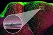
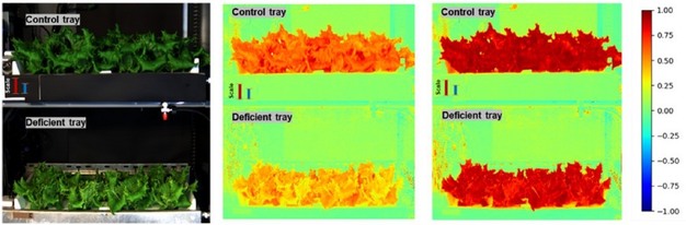 © Nanyang Technological University SingaporeFigure 2: Images derived from HSI camera can be used to distinguish lettuce heads with ideal nutrient intake (control or reference tray) and deficient nutrient intake (deficient tray)
Transforming corneal transplant assessment
One of the most immediate and impactful applications of this work lies in corneal transplantation, one of the most widely performed forms of tissue transplantation worldwide.
Currently, evaluating donor corneas relies heavily on manual inspection and contact-based microscopy. The process is time-consuming, dependent on specialist expertise and can introduce variability between assessments. Delays in evaluation can also affect how quickly patients receive sight-restoring treatment.
Dr Maria's approach offers a non-contact, data-driven alternative. By analysing spectral signatures, the system can detect subtle biochemical and structural changes within the cornea, including tissue transparency, hydration levels, collagen organisation and metabolic indicators.
These measurements provide quantitative insight into tissue quality, supporting faster and more consistent decision-making.
A more reliable and efficient evaluation process could streamline transplantation workflows and reduce waiting times for patients. For individuals awaiting vision-restoring procedures, this translates directly into improved outcomes and quality of life.
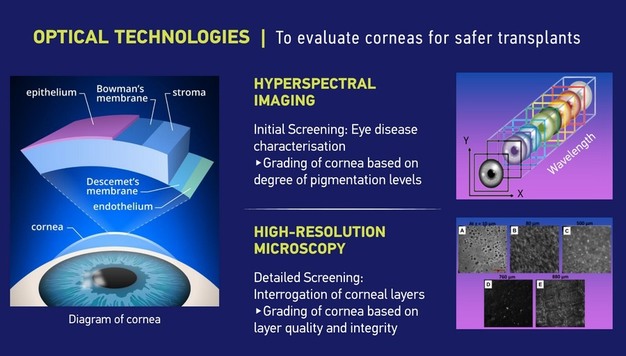 © Nanyang Technological University SingaporeFigure 3: Infographic of optical technologies used for evaluation of corneas for safer transplants
Applications across fields
While the biomedical application is a key focus, the underlying technology is highly versatile.
Dr Maria has applied hyperspectral imaging to detect early signs of stress in crops, enabling farmers to intervene before visible damage occurs. In controlled environments such as hydroponic systems, the technology can also support nutrient monitoring and optimisation, improving both yield and resource efficiency.
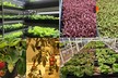
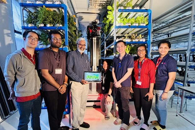 © Nanyang Technological University SingaporeDr Maria (second from right) and her team doing field trial with Urban Green Dot, a hydroponics company, as part of a project that received the Photonics Middle East Brilliance Award at the Photonics International Conference 2024 in Dubai. Prof. Murukeshan (third from the right) was the Principal Investigator of the project funded by Singapore Food Agency (SFA) and NRF Singapore.
Dr Maria has also extended her research to materials science, developing imaging approaches for corrosion monitoring, including in saline environments where degradation is difficult to detect early. These methods allow for non-invasive inspection of structural integrity, which is critical for maintaining infrastructure and ensuring safety in industrial settings.
Across these domains, the value lies in the ability to extract detailed insights quickly, accurately and without damage, supporting more informed decision-making.
Bridging lab and clinical practice
Translating these advances into real-world applications presents a distinct set of technical and practical challenges.
In clinical settings, introducing a new imaging system requires careful validation to ensure it integrates seamlessly into existing workflows. From a technical perspective, one of the main challenges is ensuring that imaging systems are robust, fast, and easy to operate and adapt in a clinical workflow.
A key challenge lies in adapting systems developed under controlled laboratory settings to less predictable clinical environments. In eye banks or hospitals, the technology must work reliably with different donor tissues, preservation conditions, and varying handling procedures.
Equally important is the development of reliable computational models. Algorithms must be trained on large and diverse datasets to ensure accuracy, consistency and generalisability in practice.
Beyond corneal applications, the concept of spectral tissue characterisation could be extended to other organs such as the kidney or liver. As each organ has different structural and biochemical properties, the spectral signatures relevant for assessing tissue health would need to be investigated, identified, and calibrated specifically for each organ type.
From research to real world impact
The next phase of Dr Maria's work focuses on clinical translation and validation.
This involves collaborating with clinical partners to test the technology on real donor tissues in a research environment. This requires careful coordination, including the establishment of Material Transfer Agreements to enable work with clinically relevant samples while ensuring ethical and regulatory compliance.
Through this collaboration, the goal is to validate the hyperspectral imaging approach on real donor tissues, compare the results with standard clinical grading methods, and refine the algorithms for clinical reliability. Such partnerships are essential because they allow laboratory-developed technologies to be tested under real eye-bank conditions.
Ultimately, these efforts could lead to the development of automated, non-contact screening tools that support clinicians in making faster and more objective decisions, improving transplantation workflows and ultimately benefiting patients awaiting treatment.